

Skin barrier function: what it is and why it matters
- Aug 10
- 11 min read
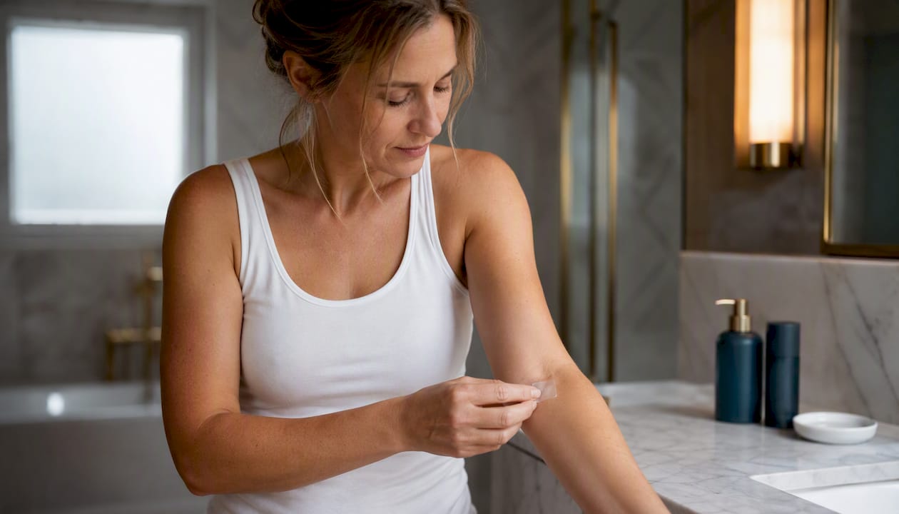
Your skin barrier is a thin but remarkably capable structure that keeps water inside your body and harmful substances out. Understanding skin barrier function comes down to two core roles: controlling transepidermal water loss (TEWL) and defending against microbes, allergens, and environmental irritants.
Sign of damage to watch for: stinging or burning when you apply products that previously felt fine.
Immediate action: simplify your routine, swap harsh cleansers for a mild, pH-balanced wash, and apply a moisturiser containing ceramides within a few minutes of cleansing.
When to seek help: if dryness, redness, or cracking persists beyond several weeks despite routine changes, or if you develop a spreading rash or signs of infection, speak to your GP.
Key takeaways
The skin barrier’s two core jobs, controlling water loss and blocking external threats, depend on an intact stratum corneum lipid matrix, a healthy acid mantle, and sufficient NMF from filaggrin breakdown.
Point | Details |
Barrier definition | The stratum corneum’s bricks-and-mortar structure controls TEWL and blocks external threats. |
Top sign of damage | Stinging with previously tolerated products signals a disrupted barrier. |
Repair routine | Layer humectant, emollient, and occlusive; choose ceramide-containing formulations. |
Key ingredient classes | Ceramides, glycerin, petrolatum, and hyaluronic acid are clinically supported for barrier repair. |
When to seek help | Persistent symptoms beyond several weeks, spreading rash, or signs of infection warrant GP review. |
Table of Contents
What is skin barrier function, exactly?
The skin barrier refers primarily to the stratum corneum (SC), the outermost layer of the epidermis. Dermatologists describe its architecture using the “bricks-and-mortar” model: flattened, protein-rich dead skin cells called corneocytes act as the bricks, held together by a mortar of intercellular lipids. Together, they regulate TEWL and block penetration of many external agents.
The key molecular players are:
Ceramides, cholesterol, and free fatty acids — the three lipid classes that form lamellar sheets in the intercellular spaces. Alterations in their composition are directly implicated in atopic dermatitis.
Filaggrin — a structural protein that breaks down into natural moisturising factor (NMF): free amino acids (a major proportion of NMF components), pyrrolidone carboxylic acid (PCA), lactate, sugars, and urea. NMF components increase humectancy and support SC recovery.
Lamellar bodies — organelles in the granular layer that store and release lipids into the intercellular space during repair.
Tight junctions — protein complexes beneath the SC that provide a secondary seal against water and solute movement.
Antimicrobial peptides (AMPs) — molecules such as defensins and cathelicidins that provide a chemical defence layer against bacteria and fungi.
Filaggrin gene mutations (common in people of Northern European ancestry, including many UK residents) reduce NMF production and are a well-established genetic risk factor for eczema and dry skin. Neonatal skin is also more alkaline and thinner, while aged skin produces fewer lipids and less NMF, making both groups more vulnerable to barrier disruption.
Pro Tip: A simple diagram of the bricks-and-mortar model is worth adding here (author to insert). Visual learners grasp the concept far more quickly when they can see how corneocytes sit within the lipid matrix.
What does the skin barrier actually do?
The barrier performs four distinct functions, and damage to any one of them tends to affect the others.
Permeability control: the lipid matrix limits water evaporation from deeper skin layers. When TEWL rises, the skin feels tight and dry, and the risk of irritant penetration increases.
Outside-in defence: the SC physically blocks microbes, allergens, and chemical irritants. AMPs add a biochemical layer, killing or neutralising pathogens before they reach living tissue.
Acid mantle: healthy skin sits at a surface pH of approximately 4.5 to 5.5. This acidity activates enzymes such as β-glucocerebrosidase and acid sphingomyelinase that generate ceramides; high-pH cleansers blunt these enzyme activities and delay barrier repair. An alkaline shift, common in newborns and elderly skin, impairs ceramide production and favours colonisation by pathogens such as Staphylococcus aureus.
Immune signalling: barrier disruption triggers cytokine release (including interleukin-1α and thymic stromal lymphopoietin), which recruits immune cells and drives the inflammatory cascade seen in eczema and contact dermatitis.
How the barrier repairs itself
Repair is surprisingly swift, but it has limits.
Minutes to hours: lamellar bodies release stored lipids into the intercellular space almost immediately after disruption, partially restoring the permeability seal. Early cytokine signalling begins recruiting repair resources.
Hours to days: lipid synthesis ramps up in the granular layer. When SC water content drops, filaggrin is broken down into NMF components, which restore humectancy and support enzyme function for desquamation. The epidermis also thickens slightly as a protective response.
Limits of repair: repeated or high-magnitude exposures, such as frequent hand washing or sustained irritant contact, can overwhelm these reparative mechanisms and prevent the SC from ever fully recovering between insults.
Pro Tip: Apply your moisturiser within a few minutes of washing while the skin is still slightly damp. This traps residual water in the SC and gives the occlusive layer something to seal in, rather than simply sitting on dry, already-depleted skin.
What damages the skin barrier?
Barrier disruption rarely has a single cause. Most people encounter several of these simultaneously.
Overwashing and harsh cleansers: high-pH soaps strip the acid mantle and remove intercellular lipids. Even water alone, applied frequently, raises skin pH and increases TEWL.
Hot showers: heat accelerates lipid extraction from the SC and temporarily raises surface pH.
Excessive exfoliation: physical scrubs and high-concentration acids remove corneocytes faster than the SC can replace them.
Low humidity and cold wind: common in UK winters, these conditions accelerate water evaporation from the SC surface.
Pollution and UV radiation: airborne particulates and UV exposure generate reactive oxygen species that degrade lipids and proteins in the SC. UV also directly damages filaggrin, reducing NMF production.
Irritant fragrances and preservatives: certain fragrance compounds and preservatives in skincare products are well-documented contact irritants that disrupt the lipid matrix.
Topical retinoids and high-strength acids: effective actives, but they accelerate cell turnover and can temporarily thin the SC during the adjustment period.
Occupational irritants: healthcare workers, hairdressers, and cleaners face repeated exposure to detergents and solvents that deplete SC lipids. These exposures increase TEWL and can produce visible dryness, fissuring, and inflammation.
Genetic factors: filaggrin mutations reduce NMF production from birth, creating a structurally weaker barrier.
Pro Tip: If hand hygiene is non-negotiable (healthcare or food-preparation work), apply a ceramide-containing hand cream immediately after each wash and keep a small tube at the sink. The few seconds it takes pays dividends in preventing fissuring.
How can you tell if your skin barrier is damaged?
Observable signs
Persistent dryness or tightness that does not resolve with standard moisturiser.
Stinging or burning when applying products you previously tolerated.
Redness, flaking, or visible fissuring.
Recurrent minor skin infections or slow-healing small wounds.
Sudden sensitivity to fragrance or preservatives in familiar products.
Clinical measurements
Clinicians and researchers use three objective tests to assess barrier status. TEWL, corneometry, and surface pH each measure a distinct property and are used in research or specialist clinics.
Measurement | What it assesses | Practical interpretation |
TEWL (transepidermal water loss) | Rate of water vapour escaping through the SC | Elevated TEWL confirms barrier disruption; used in dermatology research and specialist clinics |
Corneometry | Electrical capacitance of the SC, reflecting hydration level | Low readings indicate a dry, depleted SC; useful for tracking moisturiser efficacy |
Surface pH | Acidity of the skin surface | Values above 5.5 suggest acid mantle disruption; relevant in eczema and neonatal skin assessment |
Self-assessment is a reasonable starting point for most people. Objective testing is most useful when the diagnosis is uncertain, when monitoring treatment response in a clinical trial, or when a specialist needs to quantify severity before prescribing.
How to protect and repair your skin barrier
A layered approach works better than any single ingredient. The goal is to remove triggers, restore lipids, and lock in hydration.
Simplify cleansing: choose a mild, pH-balanced cleanser (an acidic pH). Avoid high-pH bar soaps and hot water. Lukewarm water and a gentle, fragrance-free wash are sufficient for most skin types.
Layer your moisturiser: apply in this order: humectant → emollient → occlusive.
Sun protection: a broad-spectrum SPF 30 or higher daily reduces UV-driven lipid degradation and filaggrin damage.
Environmental measures: a bedroom humidifier set to moderate relative humidity during winter during UK winter months significantly reduces overnight TEWL.
Pause actives during flares: temporarily stop retinoids and AHAs/BHAs when the barrier is visibly compromised. Reintroduce them one at a time, starting at the lowest frequency, once the barrier has stabilised.
Ingredient guidance
Moisturisers that combine ceramide-containing formulations, humectants such as glycerin and hyaluronic acid, and occlusives such as petrolatum form an evidence-based layered approach to barrier repair.
Ingredient class | Example ingredients | Primary role |
Humectant | Glycerin, hyaluronic acid, urea | Draws water into the SC from deeper layers |
Emollient | Fatty acids, squalane, shea butter | Fills gaps between corneocytes; improves texture |
Occlusive | Petrolatum, dimethicone | Seals the surface to reduce TEWL |
Lipid-replacement | Ceramides (NP, AP, EOP) | Directly replenishes the intercellular lipid matrix |
Simplifying routines, using pH-appropriate products, and choosing moisturisers with ceramides or humectants are effective initial steps for repairing a damaged barrier and reducing symptoms such as stinging, redness, and dryness. Understanding skin plumping and hydration strategies can also help you make sense of why layering works at a cellular level.
Pro Tip: During a flare, apply any prescribed topical (such as a corticosteroid) first, then your emollient on top. This sequence is consistent with NHS emollient guidance and avoids diluting the active ingredient.
Why barrier function matters for skin disease
Barrier dysfunction sits at the centre of several common dermatological conditions, not just as a consequence but as a contributing cause.
Atopic dermatitis (eczema): filaggrin mutations reduce NMF and lipid production, allowing allergens and microbes to penetrate. Changes in SC lipid composition are directly implicated in atopic dermatitis pathogenesis. Barrier impairment also increases S. aureus colonisation, which amplifies inflammation.
Contact dermatitis: a compromised barrier allows irritants and allergens to penetrate more readily, lowering the threshold for both irritant and allergic reactions.
Psoriasis: abnormal keratinocyte differentiation disrupts the lipid matrix and tight junctions, producing a leaky barrier alongside the characteristic inflammatory plaques.
Acne: disruption of the acid mantle alters the skin microbiome, potentially favouring Cutibacterium acnes overgrowth and inflammatory lesions.
The acid mantle’s disruption increases colonisation risk by pathogens such as S. aureus, particularly in newborns and elderly individuals where skin pH is naturally more alkaline. Therapeutically, this means emollient prescriptions are a first-line recommendation in atopic dermatitis management in the UK, used both to treat flares and to reduce their frequency. Targeted biologics such as dupilumab work partly by addressing the underlying barrier biology rather than simply suppressing inflammation.
When should you see a GP or dermatologist?
Most mild barrier disruption responds to routine modification within two to three weeks. Seek professional advice when:
A rash is spreading, particularly if it crosses joint creases or affects the face.
Skin cracks or fissures are bleeding or show signs of infection (warmth, pus, swelling).
Symptoms are severe, recurrent, or have not improved after four weeks of consistent barrier care.
You develop new systemic symptoms such as fever alongside a skin flare.
You suspect allergic contact dermatitis (patch testing is needed to identify the allergen).
Pro Tip: Before your GP appointment, note the onset date, any new products or occupational exposures, what you have already tried, and whether anything makes it better or worse. This information helps the clinician decide quickly whether a referral or patch testing is appropriate.
In the UK, your GP is the first port of call and can prescribe emollients, topical steroids, and referrals to NHS dermatology. Waiting times for NHS dermatology vary by region; private dermatology appointments are typically available within one to two weeks if speed is a priority.
Does diet and hydration affect your skin barrier?
Nutrition plays a supporting role in barrier health, though it works more slowly than topical care. The SC lipid matrix depends on dietary fatty acids, particularly omega-3s (found in oily fish, flaxseed, and walnuts) and omega-6s, as precursors for ceramide and fatty acid synthesis. Deficiencies in zinc, vitamin C, and vitamin D have each been associated with impaired wound healing and reduced barrier integrity, though frank deficiencies are more relevant than optimising intake in already-replete individuals.
Hydration is frequently overstated as a barrier repair tool. Drinking more water does not directly increase SC hydration in people who are already adequately hydrated; the SC gets its water from the layers below, not from the bloodstream. That said, severe dehydration does reduce skin turgor and elasticity. The more direct route to skin hydration is topical: humectants and occlusives applied to the skin surface are far more efficient at raising SC water content than increasing fluid intake.
How does skin barrier function differ across age groups and skin types?
Skin barrier competence is not fixed. It shifts across the lifespan and varies meaningfully between skin types, which matters for UK readers given the country’s diverse population and variable climate.
Neonatal skin is structurally immature at birth. The SC is thinner, surface pH is higher (closer to neutral), and TEWL is elevated compared to adult skin. This normalises over the first few weeks of life, but it means newborns are particularly vulnerable to irritants, infection, and topical absorption of substances applied to the skin.

Children and adolescents generally have a well-functioning barrier, though atopic dermatitis peaks in early childhood and is strongly associated with filaggrin mutations. Puberty brings sebaceous gland activation, which can alter the lipid profile of the SC surface.
Adult skin maintains relatively stable barrier function, though cumulative UV exposure, occupational irritants, and lifestyle factors (smoking, poor sleep, chronic stress) gradually reduce lipid production and NMF levels over time.
Aged skin (broadly, over 60) shows measurable reductions in ceramide content, filaggrin expression, and sebum production. TEWL tends to rise and corneometry readings fall, producing the characteristic dryness and fragility of older skin. This is particularly relevant in the UK, where cold, low-humidity winters compound age-related lipid depletion.
Skin type also matters. Fitzpatrick skin types I and II (common in the UK) tend to have lower melanin density, which offers less UV protection and may correlate with slightly higher baseline TEWL. Darker skin types (Fitzpatrick IV–VI) generally show higher ceramide content and lower TEWL, but are not immune to barrier disruption and may present differently (e.g. ashy appearance rather than redness) when damaged.
Our perspective on skin barrier care
The science of barrier function is well-established, yet the practical conversation around it is still dominated by single-ingredient marketing rather than the layered, trigger-removal approach the evidence supports. The most common mistake is reaching for a new active product when the barrier is already compromised. Retinoids, acids, and exfoliants all have their place, but applying them to a disrupted SC is counterproductive. Restoration comes first.
Healthy skin is also the foundation for everything else you ask of it, including how it responds to UV exposure and the natural tanning process. At NuTan®, we think about skin health holistically. Our NuTan® tanning patches work by activating the skin’s own melanocyte pathway, requiring far less UV exposure than conventional tanning methods. A well-maintained barrier means the skin is better placed to respond to that process and recover from any incidental sun exposure. The biology of natural tanning pathways and barrier care are more connected than most people realise.
Sources
This article is general information, not a substitute for advice from a qualified doctor. Consult a qualified healthcare professional about your own circumstances before acting on anything here.
FAQ
What is the skin barrier and what does it do?
The skin barrier is the outermost layer of the epidermis (stratum corneum), built from protein-rich cells and intercellular lipids. Its two core functions are preventing water loss from the body and blocking entry of microbes, allergens, and irritants.
How can you tell if your skin barrier is damaged?
The clearest sign is stinging or burning when applying products you previously tolerated without issue. Persistent dryness, redness, flaking, and recurrent minor infections also indicate barrier disruption.
What destroys the skin barrier?
Overwashing, high-pH soaps, hot water, low humidity, excessive exfoliation, irritant fragrances, UV radiation, and certain topical actives such as high-strength retinoids and acids all weaken the barrier by stripping SC lipids or disrupting the acid mantle.
How do you restore skin barrier function?
Remove the trigger first, then apply a layered moisturiser: a humectant (glycerin or hyaluronic acid), followed by an emollient, then an occlusive such as petrolatum. Choose products containing ceramides to directly replenish the intercellular lipid matrix.
When should you see a doctor about skin barrier problems?
See your GP if symptoms persist beyond four weeks of consistent barrier care, if a rash is spreading or infected, or if you suspect allergic contact dermatitis. In the UK, your GP can prescribe emollients and refer you to NHS dermatology if needed.
Recommended
Comments