

Why collagen decreases with age: the science explained
Collagen production falls with age because the body simultaneously makes less new collagen and breaks down more of what already exists, creating a net structural deficit in the skin and connective tissues. The key biological actors are dermal fibroblasts (which produce collagen), matrix metalloproteinases (MMPs, the enzymes that degrade it), transforming growth factor-β (TGF-β, the signalling molecule that drives synthesis), and reactive oxygen species (ROS, which trigger the molecular cascade that tips the balance towards breakdown). According to the Cleveland Clinic, the body produces less collagen as it ages and existing collagen degrades faster, with women commonly experiencing a marked reduction after menopause.
Clinically, this means: thinner, less elastic skin; deeper wrinkles; reduced tendon and ligament resilience; and slower wound healing.
The single highest-impact prevention step: consistent daily broad-spectrum sunscreen and avoiding smoking, both of which directly suppress the MMP activity that destroys collagen.
Key takeaways
Collagen decreases with age because fibroblast activity declines, MMP-driven degradation accelerates, and TGF-β signalling weakens, creating a net structural deficit that UV exposure, smoking, and glycation make significantly worse.
Point | Details |
Dual failure drives loss | Both reduced synthesis and accelerated MMP-driven breakdown contribute to net collagen loss with age. |
UV and smoking are the top modifiable risks | Daily broad-spectrum SPF and smoking cessation address the two highest-impact extrinsic drivers of collagen degradation. |
Retinoids have the strongest topical evidence | Retinoids modulate TGF-β signalling and suppress MMPs; expect 12–24 weeks before measurable structural improvement. |
Supplement evidence is weak | A 2025 meta-analysis of 23 RCTs found positive supplement effects concentrated in industry-funded trials; high-quality trials showed no significant benefit. |
Prevention outperforms repair | Collagen protected from further damage does not need replacing; starting protective habits early yields greater long-term benefit than later intervention. |
Table of Contents
How does collagen actually change as you age?
Collagen loss is not simply a matter of making slightly less of a protein. The structural quality of what remains also deteriorates. Fibrils become fragmented, cross-linking increases (making the matrix stiffer and more brittle), and the organised architecture that gives young skin its resilience gradually breaks down. Types I and III collagen, which together form the bulk of the dermal scaffold, are the most visibly affected.
The distinction between intrinsic and extrinsic ageing matters here, because one is unavoidable and the other is largely within your control.
Factor | Intrinsic ageing | Extrinsic ageing |
Primary drivers | Fibroblast senescence, reduced TGF-β signalling, genetic programme | UV radiation, smoking, glycation, pollution |
Typical signs | Gradual thinning, fine lines, reduced elasticity | Deeper wrinkles, uneven texture, pigmentation |
Modifiable? | No (but pace can be slowed) | Yes, substantially |
Intrinsic ageing follows a biological clock: fibroblasts become less active, signalling pathways wind down, and the ECM (extracellular matrix) loses its mechanical integrity over decades. Extrinsic ageing is superimposed on that baseline and can accelerate the process considerably, particularly through UV exposure. The practical implication is that protecting against extrinsic factors preserves the collagen you have for longer, even though the underlying biological decline cannot be stopped entirely.
Why does collagen decrease with age at the molecular level?
The cellular and molecular story behind collagen loss is more nuanced than a simple slowdown in production. It involves a self-reinforcing feedback loop between fibroblasts and the ECM they inhabit, a shift in enzyme balance, and the failure of key signalling pathways. A PMC review of dermal ageing mechanisms identifies ECM fragmentation, elevated MMP activity, and impaired TGF-β signalling as the three central drivers of net collagen loss.
Fibroblast ageing and the ECM feedback loop
Fibroblasts are the skin’s collagen factories. They require mechanical tension from an intact ECM to maintain their size, shape, and synthetic output. As collagen fibrils fragment with age, that tension drops, and fibroblasts respond by shrinking and reducing their activity. Research published in PMC shows that fibroblasts in contact with fragmented ECM produce significantly less collagen than those in contact with intact matrix, and that restoring intact ECM can partially recover fibroblast function in vitro. The fragmentation therefore creates its own momentum: less collagen leads to less tension, which leads to less collagen synthesis, which leads to more fragmentation.
The fibroblast-ECM feedback loop: fragmented collagen reduces mechanical tension on fibroblasts → fibroblasts shrink and reduce synthetic output → less new collagen is produced → existing matrix fragments further → the cycle continues. Interventions that restore ECM integrity (such as microneedling or fractional laser) aim to break this cycle by stimulating organised neocollagen.
MMP overexpression and the TIMP imbalance
MMPs are the enzymes responsible for collagen hydrolysis. MMP-1, the primary collagenase, initiates fibril fragmentation. In aged and photo-aged skin, MMP levels rise significantly, driven by ROS and the transcription factor AP-1. The problem is compounded because the endogenous inhibitors of MMPs, known as TIMPs (tissue inhibitors of metalloproteinases), do not increase proportionally. This imbalance means degradation accelerates without a matching brake. UV exposure and cigarette smoke both amplify this effect directly, pushing MMP activity higher still.
TGF-β signalling and other pathways
TGF-β is the principal growth factor that drives fibroblasts to synthesise new collagen. With age, TGF-β receptor expression and downstream Smad signalling decline, reducing the fibroblast’s response to the signals that would otherwise maintain collagen output. A PMC review on promoting collagen synthesis describes how this downregulation, alongside changes in the PI3K/Akt, JAK/STAT, and PPARβ/δ pathways, creates multiple simultaneous failures in the synthesis machinery. Hydroxylation of proline and lysine residues, a rate-limiting step for stable triple-helix formation, also becomes less efficient, meaning that even when procollagen is produced, less of it folds correctly and is secreted as functional collagen.
What environmental factors speed up collagen loss?
External exposures can dramatically accelerate the collagen depletion that intrinsic ageing would cause on its own. The Harvard Nutrition Source identifies UV exposure, smoking, and high sugar intake as the three most damaging modifiable factors, with UV ranked highest for the sheer scale of structural damage it causes.
Ultraviolet radiation (strongest evidence): UV directly induces ROS and AP-1 activation, driving MMP expression and accelerating fibril fragmentation. Chronic sun exposure produces a distinct pattern called photoageing, characterised by deep wrinkles, leathery texture, and pigmentation that goes well beyond chronological ageing. Daily broad-spectrum SPF 30 or higher is the single most evidence-supported preventive measure.
Smoking (strong evidence): Cigarette smoke generates ROS, reduces local blood flow and oxygen delivery to the dermis, and upregulates MMP activity. Smokers consistently show greater collagen loss and more pronounced wrinkling than non-smokers of the same age.
High glycaemic load and sugar (moderate evidence): Excess glucose triggers glycation, a process in which sugar molecules bind to collagen fibres and form advanced glycation end-products (AGEs). AGEs cause abnormal cross-linking that makes collagen rigid and resistant to normal remodelling. Reducing refined sugar and ultra-processed foods lowers the glycation burden on the dermal matrix.
Alcohol and poor sleep (moderate evidence): Both impair growth hormone release and cellular repair processes that occur during sleep, reducing the window for collagen synthesis and maintenance.
Pollution and chronic stress (emerging evidence): Particulate matter and cortisol from chronic stress both generate oxidative load, contributing to MMP upregulation, though the evidence base here is less mature than for UV and smoking.
Pro Tip: If you can only make one change, make it SPF. A broad-spectrum sunscreen applied every morning, regardless of cloud cover, addresses the highest-impact extrinsic driver of collagen loss. Pair it with smoking cessation for a compounding effect on MMP suppression.
It is worth noting that transdermal UV exposure (from sunlight reaching the skin) carries a substantially higher risk to collagen than incidental topical contact with skincare ingredients. The distinction matters when evaluating risk: the UV dose received during unprotected sun exposure is the primary concern, not the route of application of a product. Understanding why skin darkens after sun exposure and the UV biology behind it helps clarify why minimising UV dose is so central to collagen preservation.
How do hormones and systemic health affect collagen production?
Collagen loss is not driven by skin biology alone. Hormonal shifts and systemic health conditions alter the rate of decline in ways that go beyond what topical or lifestyle measures can fully address.
Menopause and oestrogen
Oestrogen plays a direct role in maintaining collagen synthesis. It supports TGF-β signalling, promotes fibroblast activity, and helps regulate MMP expression. After menopause, oestrogen levels fall sharply, and the skin’s collagen content can decline noticeably in the years that follow. The Cleveland Clinic notes that women commonly experience a marked reduction in collagen after menopause, and dermatologists frequently observe that the perimenopausal to post-menopausal transition is one of the most clinically visible periods of accelerated skin ageing.
Life stage callout: Collagen decline accelerates at three key points: the late twenties (when synthesis begins to slow), the early forties (when ECM fragmentation becomes more pronounced), and the perimenopausal to post-menopausal transition (when oestrogen withdrawal removes a key regulatory brake on MMP activity).
Metabolic and systemic contributors
Diabetes and insulin resistance amplify glycation, increasing AGE formation in the dermal matrix and impairing the normal collagen remodelling cycle. Chronic low-grade inflammation, common in metabolic syndrome and obesity, elevates pro-inflammatory cytokines that upregulate MMP expression. Elevated cortisol from prolonged psychological stress suppresses fibroblast activity and reduces TGF-β responsiveness, compounding the synthesis deficit. Each of these systemic factors acts on the same molecular pathways as intrinsic ageing, effectively adding years to the biological age of the dermis.
When does collagen decline start, and how fast does it fall?
Collagen synthesis begins to slow in the mid-to-late twenties for most people, though the timeline varies considerably between individuals depending on genetics, skin type, UV history, and lifestyle. By the thirties, the net balance between synthesis and degradation typically shifts into deficit. The forties tend to bring more visible structural changes as ECM fragmentation accumulates. The perimenopausal and post-menopausal years can accelerate the pace further, particularly for women.
Published numeric estimates of the rate of collagen loss vary depending on the measurement method and the population studied, so any single figure should be treated as an approximation rather than a universal rule. What the research consistently shows is that measurable molecular changes precede visible skin changes by years. The dermis begins losing structural integrity before wrinkles become apparent, which is why prevention started in the twenties and thirties has a greater impact than attempting to restore collagen architecture in the fifties or sixties.
The practical takeaway is straightforward: the earlier protective habits are established, the more collagen is preserved going into later decades. Waiting for visible signs before acting means the underlying deficit is already substantial.
What does collagen loss look like in skin and other tissues?
The clinical signs of reduced collagen are familiar, but the underlying structural changes are worth understanding because they explain why certain interventions work and others do not.
Dermatological signs:
Fine lines appearing first around the eyes and mouth, where skin is thinnest and most mobile
Deeper wrinkles as ECM fragmentation reduces the dermis’s ability to spring back after repeated movement
Reduced skin elasticity and a loss of the firm, plump texture associated with younger skin
Drier skin, partly because collagen’s hydrophilic structure contributes to water retention in the dermis
Thinning of the dermis overall, making the skin appear more translucent and fragile
Altered texture, including roughness and uneven tone, as the organised fibril architecture breaks down
Extra-dermal consequences:
Collagen is structural throughout the body, not just in skin. Tendons and ligaments rely on type I collagen for tensile strength, and reduced synthesis contributes to the slower recovery from minor injuries that many people notice from their forties onwards. Wound healing slows as the collagen scaffolding needed for tissue repair becomes less efficiently produced. Joint cartilage, which contains type II collagen, also becomes less resilient over time, contributing to the stiffness and discomfort associated with ageing joints.
A clinician assessing skin ageing will typically look for reduced recoil on a gentle pinch test (the skin takes longer to return to its original position), visible loss of jawline definition as dermal support diminishes, and the depth and distribution of wrinkles relative to the patient’s age and sun exposure history. These observations, combined with patient history, guide decisions about whether topical, procedural, or systemic approaches are most appropriate. For a closer look at how dermal volume and collagen interact with skin plumpness, skin plumping explained covers the underlying mechanics clearly.
What actually helps slow or address collagen loss?
The evidence base for collagen-related interventions ranges from very strong (daily SPF) to genuinely uncertain (oral supplements). Understanding where each option sits on that spectrum helps you build a regimen that is both realistic and grounded in science.
Prevention first
Consistent daily broad-spectrum sunscreen remains the most evidence-supported single intervention for slowing collagen loss. Smoking cessation removes one of the most potent MMP-upregulating exposures. A diet that limits refined sugar reduces glycation, while adequate vitamin C intake is non-negotiable for collagen synthesis: vitamin C is a cofactor for the prolyl and lysyl hydroxylases that stabilise the collagen triple helix, and deficiency directly impairs collagen secretion. Proline and glycine, found in bone broth and protein-rich foods, provide the amino acid substrate for procollagen assembly.
Topical agents with clinical support
Retinoids (tretinoin, retinol): The best-evidenced topical class for collagen. Retinoids modulate TGF-β signalling, suppress MMP expression, and stimulate fibroblast activity. Prescription-strength tretinoin has the strongest data; over-the-counter retinol products work through the same pathway but more slowly. Expect 12–24 weeks before structural changes become measurable.
Topical vitamin C (L-ascorbic acid): Acts as a cofactor for hydroxylation and as a direct antioxidant, reducing ROS-driven MMP activation. Stability in formulation matters: look for concentrations of 10–20% in a low-pH, anhydrous base.
Peptides: Signal peptides (such as Matrixyl/palmitoyl pentapeptide-4) aim to mimic collagen fragments and stimulate fibroblast synthesis. Evidence is promising but more limited than for retinoids or vitamin C.
Procedures with clinical evidence
Microneedling creates controlled micro-injury that triggers a wound-healing response, stimulating organised neocollagen deposition. Fractional lasers (ablative and non-ablative) work similarly, with deeper remodelling at the cost of longer recovery. Energy-based treatments such as radiofrequency and high-intensity focused ultrasound (HIFU) heat the dermis to stimulate fibroblast activity and collagen contraction. Dermal fillers do not stimulate new collagen directly but restore volume and mechanical tension in the ECM, which may secondarily support fibroblast function. Durability varies: microneedling results typically build over three to six months and require maintenance; laser remodelling can last one to two years depending on ongoing UV protection.
Oral collagen supplements: what the evidence actually says
Evidence note: A 2025 systematic review and meta-analysis of 23 randomised controlled trials (1,474 participants), published in The American Journal of Medicine, found that positive effects of collagen supplements were concentrated in industry-funded or lower-quality trials. High-quality, non-industry trials showed no significant effect on skin ageing outcomes. This does not mean supplements are harmful, but it does mean the evidence for benefit is currently weak when funding bias is controlled for.
The practical guidance from this finding is to treat oral collagen supplements as a low-certainty option rather than a cornerstone of a collagen-preservation strategy. Prevention and clinically proven topical or procedural options offer a more reliable return.
Practical regimen:
Daily: broad-spectrum SPF 30+, topical vitamin C in the morning, retinoid at night (start low, increase gradually)
Weekly: review diet for refined sugar and ensure adequate protein, vitamin C, and hydration
Clinically, when ready: consult a dermatologist or aesthetic clinician for assessment of procedural options, particularly if visible laxity or deep wrinkles are present
Pro Tip: Retinoids are the most evidence-supported topical intervention, but they require patience. Start with a low-concentration retinol two nights per week and increase frequency over eight to twelve weeks to minimise irritation. Visible improvement in texture typically appears at twelve weeks; structural collagen changes take longer.
Safety and red flags: If you notice rapid skin thinning, unusual bruising, or poor wound healing, see your GP to rule out systemic causes (including corticosteroid use, nutritional deficiency, or connective tissue conditions) before pursuing cosmetic interventions. A dermatologist referral is appropriate when over-the-counter approaches have not produced results after six months of consistent use, or when procedural options are being considered.
Can collagen decline actually be reversed?
The honest answer is: partially, and with realistic expectations. No current intervention fully restores the collagen architecture of younger skin. What the best approaches do is stimulate new collagen deposition, slow further degradation, and improve the functional quality of the existing matrix.
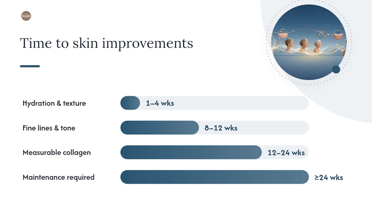
Weeks 1–4: Hydration and surface texture may improve with retinoids and vitamin C as surface cell turnover increases. No structural collagen change yet.
Weeks 8–12: Early improvements in fine lines and skin tone become visible as retinoid-driven TGF-β modulation begins to take effect.
Months 3–6: Measurable increases in dermal collagen density are possible with consistent retinoid use or after a course of microneedling or fractional laser treatment.
Months 6 and beyond: Maintenance is required to preserve gains. Without ongoing SPF, retinoid use, and UV avoidance, the same extrinsic drivers will resume their degrading effect.
Most approaches stimulate new collagen or protect the existing matrix rather than fully restoring youthful architecture. Measuring progress is best done through standardised photographs taken in consistent lighting, skin elasticity assessments using a cutometer (available in dermatology clinics), or clinician evaluation at six-month intervals. Subjective impressions of improvement are unreliable over short periods because skin appearance varies with hydration, sleep, and lighting.
The most important mindset shift is from “reversal” to “preservation and incremental improvement.” Collagen that is protected from further extrinsic damage is collagen that does not need to be replaced. Starting early, staying consistent, and managing expectations are the three principles that separate people who see genuine long-term benefit from those who cycle through products without progress.
NuTan®'s perspective on UV, skin health, and collagen
At NuTan®, skin science sits at the heart of everything we do. The evidence on collagen loss makes one thing very clear: UV exposure is among the most damaging things you can do to your skin’s structural integrity, and reducing UV dose is one of the most meaningful steps you can take for long-term skin health.
NuTan® transdermal tanning patches are designed with this principle in mind. By working with your skin’s own melanocortin pathway, the NuTan® MSH-ComplexB formula helps trigger your skin’s natural tanning response, so you can achieve a natural-looking tan with substantially less UV exposure than conventional tanning methods require. The result is a real, natural tan that does not wash or rub off, achieved through your skin’s own pigment-producing process rather than surface staining. For those who want to understand the biology behind this approach, how natural tanning pathways work explains the melanocortin signalling in detail.
If you are building a skin-health routine that takes collagen preservation seriously, pairing UV-reduction strategies with a supportive topical regimen makes sense. NuTan®'s collagen instant lift face cream is formulated to complement that approach, supporting skin appearance while you work on the longer-term structural picture.

Explore the full NuTan® range, including NuTan® tanning patches, at nutan.net. Worldwide delivery is available.
Sources
This article is general information, not a substitute for advice from a qualified doctor. Consult a qualified healthcare professional about your own circumstances before acting on anything here.
FAQ
At what age does collagen start to decline?
Collagen synthesis begins to slow in the mid-to-late twenties for most people, with the net balance between production and breakdown typically shifting into deficit during the thirties. The pace accelerates further around menopause due to falling oestrogen levels.
How can you prevent collagen loss with age?
Daily broad-spectrum sunscreen (SPF 30 or higher), smoking cessation, and a diet low in refined sugar are the three highest-impact preventive measures. Consistent use of a retinoid and topical vitamin C adds further protection by supporting TGF-β signalling and reducing MMP-driven degradation.
Is it too late to take action on collagen at 60?
No. While prevention is more effective than repair, retinoids, topical vitamin C, and procedural options such as microneedling and fractional laser can still stimulate measurable new collagen deposition at any age. A dermatologist can assess which options are most appropriate for your skin’s current condition.
Can collagen decline be reversed?
Fully restoring youthful collagen architecture is not currently possible, but consistent use of retinoids, procedural treatments, and rigorous UV protection can stimulate new collagen deposition and slow further loss, producing genuine improvements in skin texture, elasticity, and firmness over three to six months.
Do collagen supplements work?
A 2025 meta-analysis of 23 randomised controlled trials found that positive effects from oral collagen supplements were concentrated in industry-funded or lower-quality studies. High-quality, independently funded trials showed no significant effect, making supplements a low-certainty option compared with proven topical and procedural approaches.
Recommended
Comments